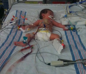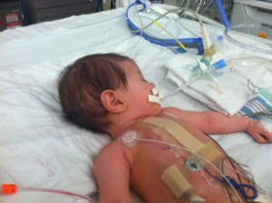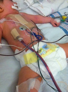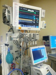
Cori Lou is out of surgery and being monitored through a very carefully managed recovery. Open heart surgery was much more invasive and disruptive than the abdominal surgery. Still, she seems to be doing well for a little baby who has just gone through what she has. (click on any photo to enlarge)
Here is what they ended up fixing:
- The ventricular septal defect (VSD) was patched, meaning the opening between the two larger chambers of her heart.
- An atrial septal defect (ASD) was patched, which you are probably guessing is an opening between the two small, or upper, chambers of her heart. This is the hole that is normal for babies to have when they are still in the womb, since their blood isn’t traveling through their lungs yet. It is only a defect when it doesn’t close up soon after birth. In conjunction with another septal defect, you have all kinds of ineffective blood flow.
- The patent ductus arteriosus (PDA) was “clipped:. This is another normal heart feature in the womb, being a vessel that connects the vessels going to and from the lungs from the heart, thus bypassing the lungs themselves since the placenta is the baby’s source of oxygen.

There was also what else they found:
- The veins transporting the blood from the liver (hepatic veins) attached directly to the heart, instead of attaching to the inferior vena cava (inferior meaning coming from the lower parts of the body), a main vein that many other veins connect up to. This, along with her extra superior vena cava meant there were lots of vessels to work around.
- Her VSD was in an awkward location, making the surgery take the full 6 hours.
- Her liver looked very normal to the surgeon, who could not see anything indicating it hadn’t always been there right where it is supposed to be.
What is going on in recovery right now:
- She is paralyzed to keep her quiet and limit any strain on the heart, for at least the first night. There were some early signs of the heart trying to force the wrong sort of rhythms, but this is somewhat expected for young babies within the first week after surgery. It is kept under control with the paralyzation and restricted movement.
- Last night, she did have some trouble with what is called a junctional rhythm. This means that the electrical junctions between the atria and ventricles (smaller and larger chambers of the heart) is not working smoothly. From what I read, this means that the heart is trying to “fix” what it perceives as too slow of a beat, but it makes the pumping of the chambers out of sync and thus not as effective. The pacemakers wires mentioned below were used to counteract this.
- She is on a ventilator. They will consider weaning her off of it as the paralyzation wears off and she shows signs of breathing on her own.
- She had a blood transfusion, which is normal for this surgery.
- There is some swelling, particularly in the chest area. This is normal after surgery.

She has many tubes in and wires on her right now:
- There is a big chest tube to keep the surgery site drained.
- Another nasogastric tube (through the nose to the stomach, like she had in the NICU after her abdominal surgery) to keep her stomach clear. (not in photo)
- A red arterial line for drawing blood and check circulatory pressures. Arterial meaning that one end is placed in an artery.
- A clear atrial line for giving specific medications. Atrial meaning one end is placed directly into an atrium (small heart chamber)
- More than one IV, for things like fluids (she isn’t eating by mouth right now again), pain medicines, and antibiotics.
- The ventilator tube is going through her mouth. For the sake of clarity, the ventilator is a machine that controls the air in and out of her lungs automatically, at a rate that is close to what her body would do.
- The glowing red spot on her left foot is her oxygen saturation monitor, keeping track of whether or not there is enough oxygen in her blood. It is not invasive, but is held against the skin.
- The tube coming out from under her diaper is draining her bladder. It is called a Foley catheter. There is some blood, which is considered normal after the body has been managed with the pressures of the heart-lung machine.
- Coiled wires on her chest are pacemaker wires, available to stimulate her heart with corrective electrical signals as needed.
- The round, white sticky pads with attached wires are the ubiquitous electrocardiogram (EKG) monitor nodes, to keep track of her heart rate and patterns.
- A rectal thermometer is closely measuring her body temperature, which they are keeping on the low side in order to keep her metabolism slow for now.
The larger bandage down the center of her chest is covering the main surgery site. There is another bandage, lower and to her right, over where the drainage tube is inserted. And another smaller on over the point of entry of the pacemaker wires. The tape around her mouth is to keep the ventilator tube secure. There is tape and such any place there is an IV. The boards on her one foot and one arm are to restrict movement of those areas so that IVs and monitors stay in place and reliable.
All the information from the medical staff, as well as things I read as I research to make sure I am presenting this as well as possible, indicate that this surgery has an almost 100% success rate in relatively uncomplicated situations such as Cori’s. Cori Lou is not a statistic, but in this case the statistics are quite encouraging. It may be time for a little beauty sleep right now, but we expect to see Cori Lou’s bright eyes again soon.


praying for your sweetie
Laura,
I so appreciate your clear and concise descriptions! I actually understand everything you wrote. Well done. 🙂 This little gal has gone through so very much and yet is so alert. She’s such a beautiful blessing! I’ll most definitely keep Cori Lou on my ongoing prayer list~and her momma and daddy. I look forward to reading her updates and seeing her sweet pictures! God bless, friend.
Thanks, Bonnie. It helps to hear from people that the reports are understandable and being read.